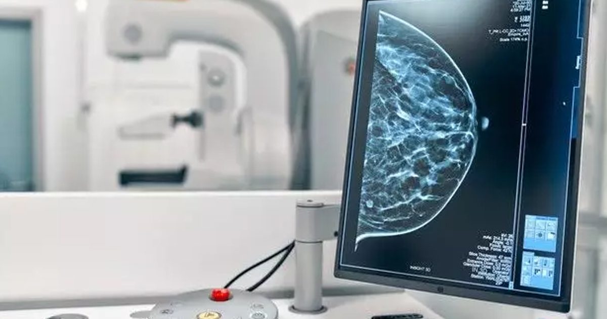
Researchers at Duke University created an Artificial Intelligence (AI) platform that would be capable of detecting and analyzing lesions in mammography images. Its objective is to be a valuable support in the decision-making of professionals.
Researchers at Duke University in Durham, North Carolina, suggest that this platform can determine if patients require biopsies if the lesions detected are potentially cancerous. The AI algorithm was trained on 1,136 images taken from 484 patients in the University Health System.
The team from the aforementioned university trained the platform's algorithm to detect and evaluate lesions as if it were a real radiologist. Unlike other algorithms that are freely trained in their procedures.
“If a computer is going to help make important medical decisions, doctors need to trust that the AI is basing its conclusions on something that makes sense. We need algorithms that not only work, but are self-explanatory and show examples of what they are basing their conclusions on. That way, whether a doctor agrees with the result or not, the AI is helping make better decisions," said Joseph Lo, a professor of radiology at Duke.
In the press release from Duke University, they explained that the algorithms that read medical images represent a large industry, since the Food and Drug Administration (FDA) has approved more than 100 for clinical use. However, there is a lot of room for improvement especially to use larger validation data sets, especially when it comes to medical images, or to include demographic information.
“Our idea was instead to build a system to say that this specific part of a possible cancerous lesion looks a lot like this one I've seen before,” said Alina Barnett, a PhD in computer science at Duke and first author of the study.
In this sense, the AI system, proposed by the university, does not seek to replace the specialist's experience, but to be a support so that professionals have more information for decision making.
The Duke University study was published in the journal Nature Machine Intelligence in December 2021: https://www.nature.com/articles/s42256-021-00423-x
DUKE UNIVERSITY
https://pratt.duke.edu/about/news/ai-breast-cancer
HEALTH IT ANALYTICS
https://healthitanalytics.com/news/ai-detectscancerouslesions-in-mammography-scans