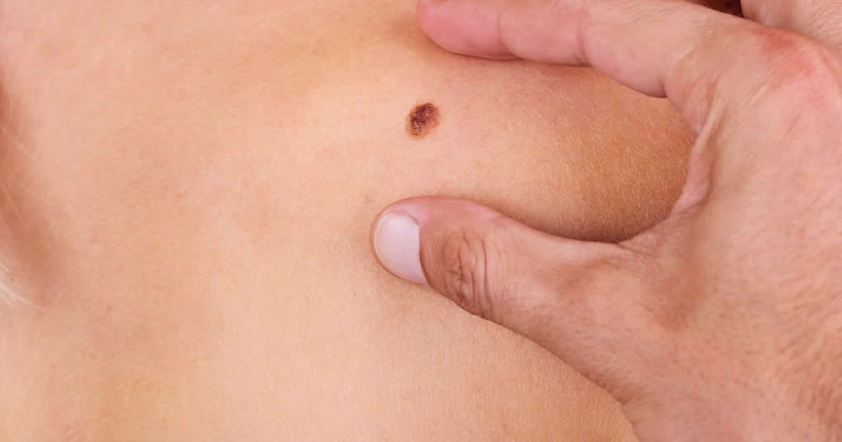
Dermatological researchers and specialists in engineering and Artificial Intelligence (AI), developed an algorithm through a convolutional neural network to examine skin lesions, through photographs, even mobile phone photos.
In the United States in 2019 more than 96,000 people were diagnosed with melanoma, a disease, 7,230 people died that year. The process of early identification of suspicious pigmented lesions (SPL) is important to reduce the cost of treatment by up to 20 times. However, the number of efficient tools for SPL detection is still limited in a country like the United States. That is why the researchers developed this algorithm to improve diagnosis.
The results were published in Science Translational Medicine under the title: “Using deep learning for dermatologist-level detection of suspicious pigmented skin lesions from wide-field images.”
MIT, Harvard and other institutions researchers built a deep learning-based algorithm for evaluating skin lesions and classifying them to rule out diseases such as skin cancer. “Rather than evaluate a single lesion at a time looking for predetermined signs of neoplasia, the algorithm identifies lesions that differ from most of the other marks on that patient’s skin, flagging them for further examination and ranking them in order of suspiciousness. The algorithm performed similarly to board-certified dermatologists and could potentially be used at primary care visits to help clinicians triage suspicious lesions for follow-up.”
The algorithm's operation is based on deep convoluted neural networks DCNNs, which through analysis of wide-field photographs is able to obtain large samples of skin sections for diagnosing diseases before they develop.
38,283 dermatological data from 133 patients and other publicly available images were collected for the study. “Our system achieved more than 90.3% sensitivity (95% confidence interval, 90 to 90.6) and 89.9% specificity (89.6 to 90.2%) in distinguishing SPLs from nonsuspicious lesions, skin, and complex backgrounds, avoiding the need for cumbersome individual lesion imaging,” the study explains.
MIT researchers contacted in 2014 the Spanish physician, Dr. José Avilés Izquierdo, from the Hospital General Universitario Gregorio Mañón in Madrid, -who is one of the co-authors of the study published in the journal Science- for the development of a machine learningproject for the detection of malignant melanomas. According to Dr. Avilés Izquierdo, the purpose of the study was "to develop an algorithm that would serve as an evaluation platform for different physical and computer tools aimed at the early diagnosis of skin cancer, especially melanoma.
Dr. Avilés Izquierdo reported his experience on the specialized site Medscape: "The researchers at the Massachusetts Institute of Technology developed a system for analyzing images of suspicious pigmented lesions based on 399 variables related to symmetry, borders, colors, texture or size. They analyzed 38,283 images from patients and public resources (image banks, atlases, and search engines)".
You can find the link to the study at the following link: https://stm.sciencemag.org/content/13/581/eabb3652